


As part of the European Nanopathology Project (QOL-147-2002-05) carried out in collaboration with the universities of Mainz and Cambridge as well as with FEI, a new electron-microscopy technique on biological sections was devised. Through that technique, the presence of micro-and nanosized foreign bodies in the tissues can be detected and, in fact, in each matrix. For the first time we can realize beyond any possible doubts that particles ranging in size from a few microns down to a few tens of nanometers (a micron is a thousandth of a millimeter and a nanometer is a thousandth of a micron) can travel in the human body carried by the blood, be imprisoned by tissues and trigger biological reactions that can be lethal.
The technique, then perfected and extended thanks to the European project of nanotoxicology DIPNA (NMP, 02321, 2006-09), can be applied to pathological sections embedded in paraffin blocks containing the pathological tissue, but it is also possible to work on fresh and mummified tissues and on fluids. Human or animal tissue, in fact, is degraded but particles do not disappear, remaining unchanged forever.
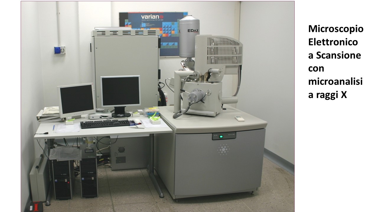
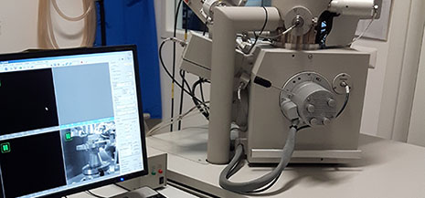
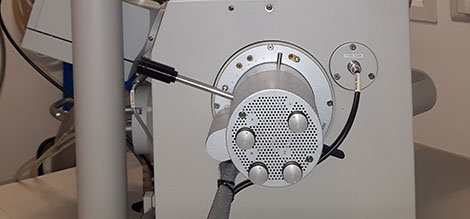
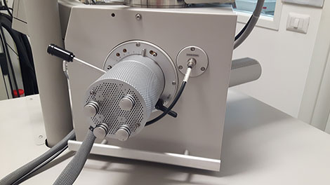
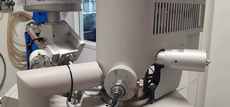
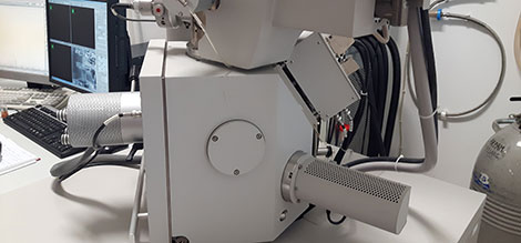
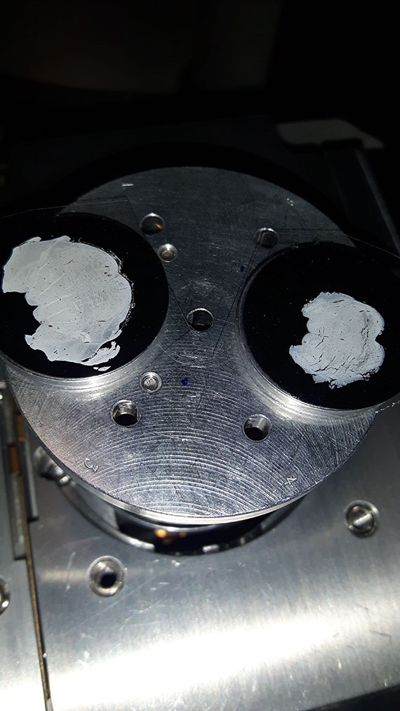
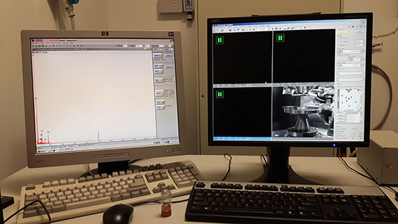
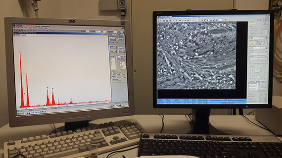
The sections of the prepared pathological tissue are deposited on an aluminum support and inserted into a scanning-electron microscope, better if the model has a so-called field-emission cannon. The equipment is completed with an X-ray microanalysis (energy-dispersion spectroscopy) to identify the elemental chemical composition of the particles detected.
Very interesting results are also obtained by analyzing many other matrices such as, for example, food, drugs, cosmetics and environmental findings such as air, water, soil, etc.